

Ultrasound
Ultrasound is an extremely valuable diagnostic in equine medicine and lameness as it is a non-invasive technique that provides real-time images and information. Dr. Renier offers ultrasound examinations of the horse’s abdomen, chest, heart, eye, and legs. Transabdominal evaluation of pregnancies is also offered.
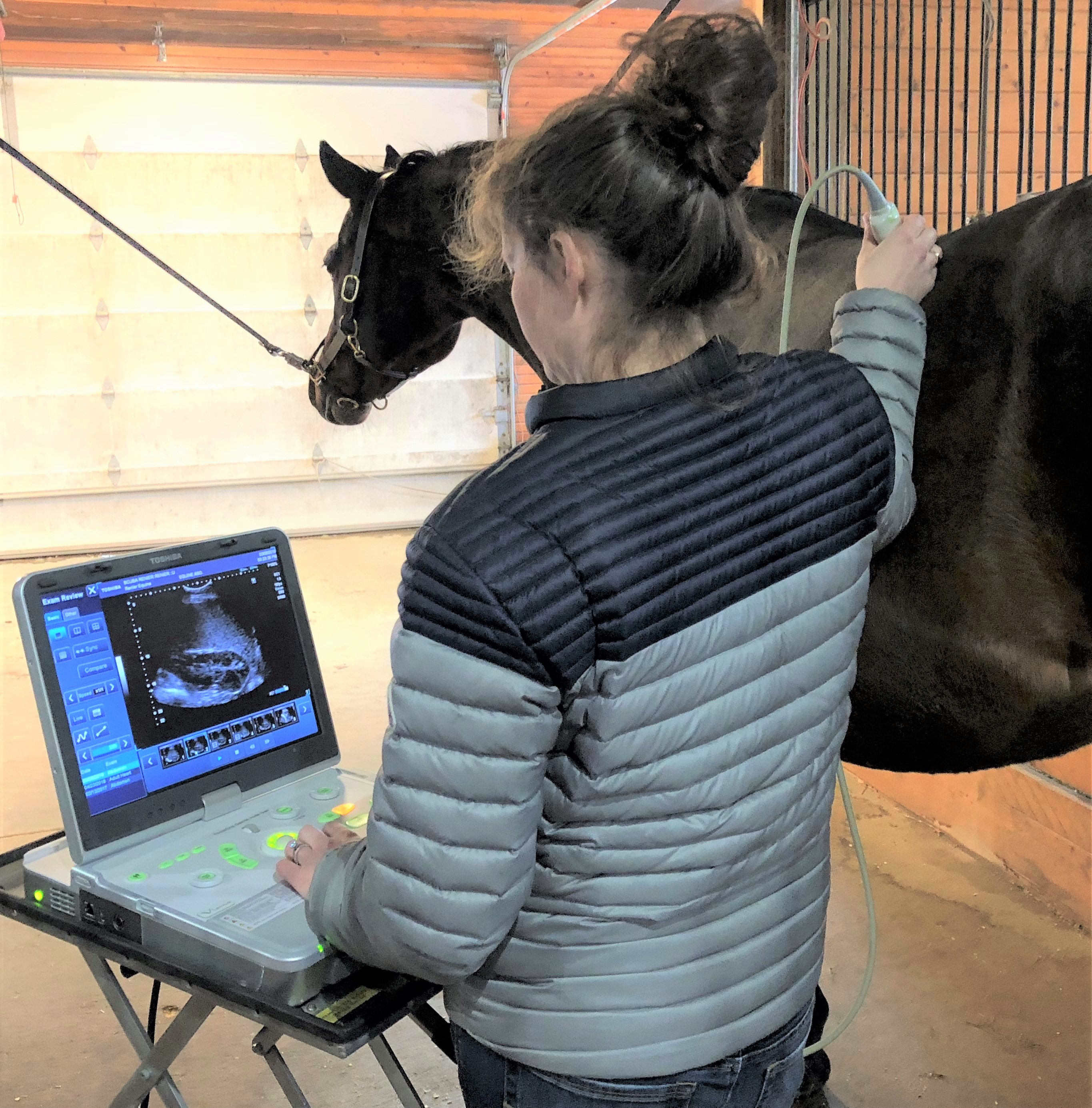
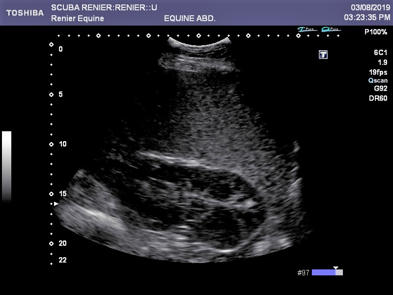
Ultrasound can be useful in both emergency situations and in diagnosing more chronic conditions. By using sound waves, ultrasound can safely, non-invasively show us the condition of internal organs. In emergency situations, such as in colic, this allows us to evaluate the activity, diameter, and thickness of parts of the intestine and to check for fluid in the abdominal cavity. Organs such as the spleen, kidneys, and liver can also be evaluated. Ultrasound of the chest cavity can show when free fluid is present around the lungs and heart or when there is an abnormality on the lung surface. Ultrasound can also show us changes in the lens of the eye, valves and chambers of the heart, and can help identify and monitor infections in a foal’s umbilicus. In orthopedic injury, ultrasound is the mainstay of soft tissue imaging on the farm. Injuries to the suspensory ligament, superficial digital flexor tendon, and deep digital flexor tendon may be visualized on ultrasound of the limb, among other conditions.